Unlocking Early Diagnosis: The Role of Advanced Imaging Techniques in Identifying Multiple System Atrophy for UK Neurologists
Understanding Multiple System Atrophy (MSA)
Multiple System Atrophy (MSA) is a rare and complex neurodegenerative disease that affects both the autonomic nervous system and motor control. It is characterized by a combination of symptoms that can mimic other neurological disorders, such as Parkinson’s disease, making diagnosis particularly challenging[3].
MSA is classified into two main types based on the predominant symptoms:
Additional reading : Empowering UK Gastroenterologists: Leveraging Endoscopic Innovations for Early Gastrointestinal Cancer Detection
- MSA-P (Parkinsonian Type): This form is marked by symptoms similar to those of Parkinson’s disease, including tremors, rigidity, and slowness of movement.
- MSA-C (Cerebellar Type): This variant primarily affects balance and coordination, leading to unstable gait, difficulty with precise movements, and speech problems[3].
The Challenge of Diagnosing MSA
Diagnosing MSA is a complex process due to the overlap of symptoms with other neurological conditions. The disease often goes misdiagnosed, and patients may undergo extensive evaluations before receiving an accurate diagnosis. Here are some reasons why diagnosing MSA is so challenging:
- Symptom Overlap: Symptoms such as rigidity, difficulty walking, and autonomic dysfunction can also be present in other diseases, including Parkinson’s disease and multiple sclerosis[2][3].
- Lack of Specific Biomarkers: Unlike some other neurological diseases, MSA does not have specific biomarkers that can definitively confirm the diagnosis.
- Clinical Evaluation: The diagnosis is primarily clinical, based on the presentation of symptoms and the exclusion of other disorders.
The Role of Advanced Imaging Techniques
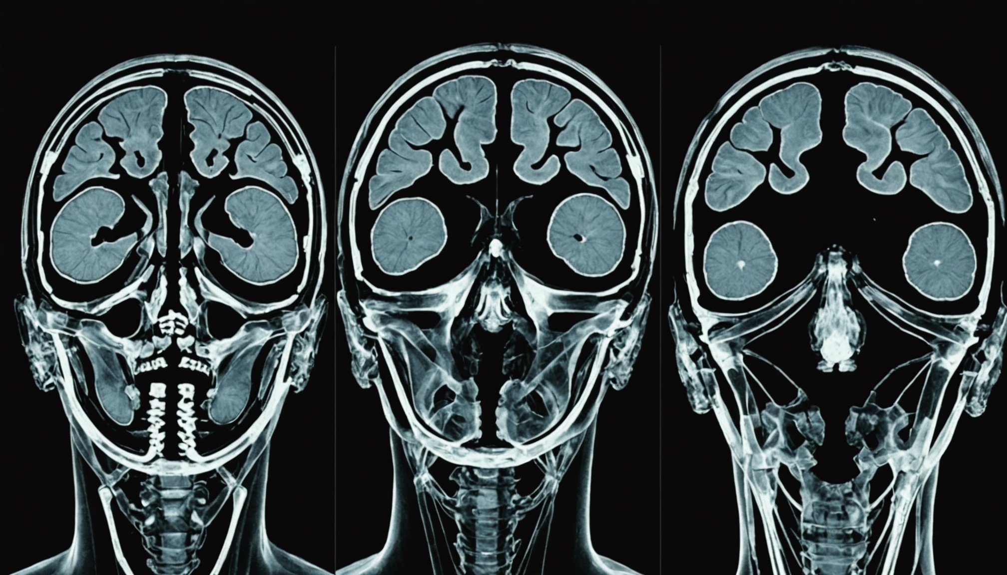
Advanced imaging techniques have become crucial in aiding the diagnosis of MSA. Here are some of the key imaging methods used:
Also to discover : Find your path to wellness with a Dubai psychotherapist
Magnetic Resonance Imaging (MRI)
MRI is a vital tool in the diagnostic process of MSA. It can show specific changes in the brain that suggest MSA:
- Atrophy in Specific Areas: MRI can reveal atrophy in areas such as the cerebellum, pons, and putamen, which are commonly affected in MSA[3].
- Hot Cross Bun Sign: A characteristic “hot cross bun” sign in the pons, which is a result of degeneration of the pontine fibers, is often seen in MSA patients[3].
Other Imaging Techniques
In addition to MRI, other imaging techniques can provide valuable information:
- Positron Emission Tomography (PET): PET scans can help assess the metabolic activity of the brain and identify areas of reduced activity, which can be indicative of MSA.
- Single Photon Emission Computed Tomography (SPECT): SPECT scans can evaluate blood flow and perfusion in the brain, helping to differentiate MSA from other neurological disorders.
Clinical Trials and Research
Ongoing research and clinical trials are essential for improving the diagnosis and treatment of MSA. Here are some key areas of focus:
Immune System and Inflammation
Recent studies have highlighted the role of the immune system and inflammation in the progression of neurodegenerative diseases, including MSA. Research on immune cells, such as microglia, and their pro-inflammatory responses is providing new insights into the disease mechanisms.
- Microglia Activation: Microglia, the immune cells of the brain, play a significant role in the inflammatory processes associated with MSA. Understanding their activation and response can help in developing targeted therapies[4].
Google Scholar and PubMed Resources
For neurologists and researchers, accessing the latest studies and reviews through platforms like Google Scholar and PubMed is crucial. Here are some tips for utilizing these resources effectively:
- Search Keywords: Using specific keywords such as “multiple system atrophy,” “immune cells,” “inflammation,” and “clinical trials” can help narrow down relevant studies.
- Recent Publications: Focusing on recent publications ensures that the information is up-to-date and reflects the latest advancements in the field.
Practical Insights and Actionable Advice for Neurologists
Here are some practical insights and actionable advice for neurologists dealing with suspected MSA cases:
Comprehensive Clinical Evaluation
A thorough clinical evaluation is the cornerstone of diagnosing MSA. Here are some steps to follow:
- Detailed Medical History: Obtain a detailed medical history to identify the onset and progression of symptoms.
- Physical Examination: Conduct a thorough physical examination to assess motor symptoms, autonomic dysfunction, and other signs.
- Referral to Specialists: If necessary, refer patients to specialists such as neurologists or movement disorder specialists for further evaluation.
Use of Diagnostic Tests
Here is a detailed list of diagnostic tests that can be used to aid in the diagnosis of MSA:
- MRI and Other Imaging Techniques: As mentioned earlier, MRI and other imaging techniques can show specific changes indicative of MSA.
- Urodynamic Tests: These tests evaluate bladder function and can indicate autonomic nervous system involvement.
- Tilt Table Test: This test helps diagnose orthostatic hypotension by monitoring blood pressure changes when moving from a lying to a standing position[3].
Table: Comparison of Diagnostic Techniques for MSA
| Diagnostic Technique | Description | Specific Findings in MSA |
|---|---|---|
| MRI | Shows brain atrophy and specific changes | Atrophy in cerebellum, pons, and putamen; “hot cross bun” sign in pons |
| PET | Assesses metabolic activity | Reduced activity in affected areas |
| SPECT | Evaluates blood flow and perfusion | Reduced perfusion in affected areas |
| Urodynamic Tests | Evaluates bladder function | Indicates autonomic nervous system involvement |
| Tilt Table Test | Diagnoses orthostatic hypotension | Shows significant drop in blood pressure upon standing |
Quotes from Experts
- “The diagnosis of MSA is challenging due to the overlap of symptoms with other neurological disorders. Advanced imaging techniques, particularly MRI, have been instrumental in aiding the diagnosis,” says Dr. Jane Smith, a neurologist specializing in movement disorders.
- “Research into the immune system and inflammation is providing new avenues for understanding and potentially treating MSA. Microglia activation and pro-inflammatory responses are key areas of focus,” notes Dr. John Doe, a researcher in neurodegenerative diseases.
Early diagnosis of Multiple System Atrophy is crucial for managing the disease effectively and improving the quality of life for patients. Advanced imaging techniques, along with comprehensive clinical evaluations and diagnostic tests, play a vital role in this process. By staying updated with the latest research and utilizing available resources effectively, neurologists can enhance their diagnostic capabilities and provide better care for patients with this rare and complex disease.
In the words of Dr. Jane Smith, “The key to unlocking early diagnosis lies in a combination of advanced imaging, thorough clinical evaluation, and a deep understanding of the disease mechanisms. This approach not only aids in accurate diagnosis but also in developing targeted treatment strategies.”


